Galactose Binding-Soluble 3 ELISA Kit
Goose Lectin-Galactose Binding-Soluble three ELISA Equipment from MYBIOSOURCE INC.
| Product Identify | Goose Lectin-Galactose Binding-Soluble three ELISA Equipment |
|---|---|
| Description | Assay Sort: Sandwich. |
| Measurement | n/a |
| Focus | n/a |
| Purposes | n/a |
| Different Names | [LGALS3; LGALS3 protein; galectin-3; lectin L-29; 35 kDa lectin; MAC-2 antigen; IgE-binding protein; laminin-binding protein; galactose-specific lectin 3; carbohydrate-binding protein 35; lectin, galactoside-binding, soluble, 3], [LGALS3; LGALS3; L31; GAL3; MAC2; CBP35; GALBP; GALIG; LGALS2] |
| Gene, Accession, CAS # | [LGALS3], Gene ID: 3958, NCBI: CAG46894.1, UniProt: Q6FGL0 |
| Catalog # | MBS018373 |
Antibodies
We’re dedicated to elevating all of our antibodies in-house. We are able to hint again every lot again to the unique host it was raised from. We even synthesize the antigens used to boost our antibodies. No extra questioning whether or not the antibody you might be utilizing is simply being re-sold below a special label.
Rat Lectin-galactose binding-soluble three ELISA Equipment
| Producer | MyBioSource |
|---|---|
| Class | ELISA |
| Sort | Elisa-Equipment |
| Particular in opposition to | Rat |
| Quantity | 96-Strip-Wells |
| Merchandise no. | MBS722004-96 |
| eClass 6.1 | 32160605 |
| eClass 9.0 | 32160605 |
| Sensitivity |
1.Zero pg/mL.
|
| Samples |
Serum, plasma, Cell Tradition Supernatants, physique fluid and tissue homogenate
|
| Gene Names |
Lgals3 ; L31; GAL3; MAC2; CBP35; GALBP; GALIG; LGALS2
|
| Assay Sort |
Sandwich
|
| Host/Reactivities |
Reactivity: Rat
|
Affinity® ECL Western Blotting Substrate (fg grade)
Introduction
The Affinity ECL Western Blotting Substrate is a extremely delicate nonradioactive, enhanced luminol-based chemiluminescent substrate for the detection of horseradish peroxidase (HRP) on immunoblots. Affinity ECL Western Blotting Substrate permits the detection of femtogram quantities of antigen and permits for straightforward detection of HRP utilizing photographic or different imaging strategies. Blots may be repeatedly uncovered to X-ray movie to acquire optimum outcomes or stripped of the immunodetection reagents and re-probed. The particular formulation of Affinity ECL Substrate makes it the best substitute for different ECL Substrate with out the necessity for extra optimization of assay circumstances.
Essential Product Data
- AffinityECL Substrate requires extra dilute antibody concentrations than these used with precipitating colorimetric HRP substrates. To optimize antibody concentrations, carry out a scientific dot blot evaluation.
- Empirical testing is important to find out the suitable blocking reagent for every Western blot system, as crossreactivity of the blocking reagent with antibodies causes nonspecific sign. Blocking buffer additionally impacts system sensitivity.
- Keep away from utilizing milk as a blocking reagent when utilizing avidin/biotin methods as a result of milk comprises variable quantities of endogenous biotin, which causes excessive background sign.
- Use adequate volumes of wash buffer, blocking buffer, antibody answer and substrate working answer to cowl blot and be sure that it by no means turns into dry. Utilizing giant blocking and wash buffer volumes minimizes nonspecific sign.
- For optimum outcomes, use a shaking platform throughout incubation steps.
- Add Tween™-20 Detergent (closing focus of 0.05-0.1%) to the blocking buffer and all diluted antibody options to attenuate nonspecific sign.
- Don’t use sodium azide as a preservative for buffers. Sodium azide is an inhibitor of HRP and will intervene with this technique.
- Don’t deal with membrane with naked fingers. At all times put on gloves or use clear forceps.
- All tools have to be clear and freed from overseas materials. Metallic gadgets (e.g., scissors) should have no seen indicators of rust. Rust might trigger speckling and excessive background.
- Publicity to the solar or another intense gentle can hurt the substrate. For greatest outcomes maintain the substrate working answer in an amber bottle and keep away from extended publicity to any intense gentle. Quick-term publicity to typical laboratory lighting won’t hurt the working answer.
Process Abstract
Observe: Antigen and antibody quantities might require optimization.
- Dilute the first antibody to5.0-0.5µg/mL.
- Dilute the secondary antibody to 1.0-0.067µg/mL.
- Combine Detection Reagents 1 and a pair of at a 1:1 ratio and add it to the blot. Incubate blot for 1-60 seconds.
- Drain extra reagent. Cowl blot with a transparent plastic sheet protector or clear plastic wrap.
- Expose blot to X-ray movie.
Detailed Western Blotting Process
- Take away blot from the switch equipment and block nonspecific websites with Blocking Reagent for 60 minutes at room temperature (RT) with shaking. If desired, block in a single day at 2-8ºC with out shaking.
- Take away the Blocking Reagent and add the first antibody working dilution. Incubate blot for 1 hour at RT with shaking or in a single day at 2-8°C with out shaking.
- Briefly rinse membrane in Wash Buffer two occasions.
- Wash membrane by suspending it in Wash Buffer and agitating for ≥ 5 minutes. Substitute Wash Buffer not less than 4-6 occasions. Rising the Wash Buffer quantity, the variety of washes and wash period might assist decrease background sign.
- Incubate blot with the HRP-conjugate working dilution for 1 hour at RT with shaking.
- Repeat Steps three and Four to take away nonbound HRP-conjugate.
Observe: Membrane MUST be completely washed after incubation with the HRP-conjugate.
- Put together the substrate working answer by mixing equal components of Detection Reagents 1 and a pair of. Use 0.125mL Working Answer per cm2 of membrane.
Observe: For greatest outcomes put together working answer instantly earlier than use. The working answer is steady for 1 hour at RT.
- Incubate blot with working answer for 1 minute at RT.
- Take away blot from working answer and place it in a plastic sheet protector or clear plastic wrap. Use an absorbent tissue to take away extra liquid and to fastidiously press out any bubbles from between the blot and the membrane protector.
- Place the protected membrane in a movie cassette with the protein aspect dealing with up. Flip off all lights besides these applicable for X-ray movie publicity (e.g., a crimson safelight).
Observe: Movie should stay dry throughout publicity. For optimum outcomes, carry out the next precautions:
- Be sure that extra substrate is faraway from the membrane and the membrane protector.
- Use gloves throughout the complete film-handling course of.
- By no means place a blot on developed movie, as chemical substances on the movie might cut back sign.
- Fastidiously place X-ray movie on prime of the membrane. Carry out a first-time publicity of30 seconds. Range the publicity time to realize optimum outcomes.
- Develop movie utilizing applicable growing answer and fixative. If sign is simply too intense, cut back publicity time or strip and re-probe the blot with decreased antibody concentrations.
Storage
Upon receipt retailer at room temperature.
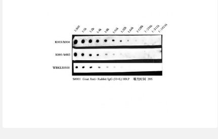
| Cat.# | Product Identify | Measurement |
| Okay003 | Answer Ⅰ | 25/50/250 ml |
| Okay004 | Answer Ⅱ | 25/50/250 ml |
 Anti-IL-13 Antibody, Mouse Monoclonal |
|||
| MBS8102506-01mL | MyBiosource | 0.1mL | EUR 300 |
Anti-IL-13 Antibody, Mouse Monoclonal |
|||
| MBS8102506-5x01mL | MyBiosource | 5x0.1mL | EUR 1200 |
Anti-IL-13 Antibody, Mouse Monoclonal |
|||
| MBS8102507-01mL | MyBiosource | 0.1mL | EUR 300 |
Anti-IL-13 Antibody, Mouse Monoclonal |
|||
| MBS8102507-5x01mL | MyBiosource | 5x0.1mL | EUR 1200 |
Anti-IL-13 Antibody, Mouse Monoclonal |
|||
| MBS8102510-002mL | MyBiosource | 0.02mL | EUR 175 |
Anti-IL-13 Antibody, Mouse Monoclonal |
|||
| MBS8102510-01mL | MyBiosource | 0.1mL | EUR 320 |
Anti-IL-13 Antibody, Mouse Monoclonal |
|||
| MBS8102510-5x01mL | MyBiosource | 5x0.1mL | EUR 1300 |
 Mouse Monoclonal Anti-IL-13 Antibody |
|||
| TA352743 | Origene Technologies GmbH | 500 µg | Ask for price |
 Mouse Monoclonal anti-human IL-13 |
|||
| hAP-0365 | Angio Proteomie | 100ug | EUR 250 |
, Mouse Monoclonal) Anti-IL-13 Antibody (HRP), Mouse Monoclonal |
|||
| MBS8102509-01mg | MyBiosource | 0.1mg | EUR 345 |
Anti-IL-13 Antibody (HRP), Mouse Monoclonal |
|||
| MBS8102509-5x01mg | MyBiosource | 5x0.1mg | EUR 1405 |
 IL-13, Mouse |
|||
| E34M036M | EnoGene | 5 μg | EUR 155 |
 Mouse IL-13 |
|||
| MBS691604-0002mg | MyBiosource | 0.002mg | EUR 300 |
Mouse IL-13 |
|||
| MBS691604-001mg | MyBiosource | 0.01mg | EUR 450 |
Mouse IL-13 |
|||
| MBS691604-5x001mg | MyBiosource | 5x0.01mg | EUR 1725 |
 IL-13, Mouse |
|||
| MBS8575363-0005mg | MyBiosource | 0.005mg | EUR 245 |
IL-13, Mouse |
|||
| MBS8575363-5x0005mg | MyBiosource | 5x0.005mg | EUR 940 |
, Mouse Monoclonal) Anti-IL-13 Antibody (Biotin), Mouse Monoclonal |
|||
| MBS8102508-01mg | MyBiosource | 0.1mg | EUR 345 |
Anti-IL-13 Antibody (Biotin), Mouse Monoclonal |
|||
| MBS8102508-5x01mg | MyBiosource | 5x0.1mg | EUR 1405 |
, Antibody) IL-13/Interleukin-13 (mouse), Antibody |
|||
| GWB-81CEEF | GenWay Biotech | 1 ml | Ask for price |
 Interleukin-13 (IL-13 Mouse |
|||
| GWB-FB2D79 | GenWay Biotech | 0.01 mg | Ask for price |
 mAb mouse anti-monkey IL-13 Cy5-labeled |
|||
| CT119 | U-CyTech | 50 µg | EUR 312 |
 mAb mouse anti-human IL-13 FITC-labeled |
|||
| CT246 | U-CyTech | 50 µg | EUR 312 |
 mAb mouse anti-monkey IL-13 FITC-labeled |
|||
| CT120 | U-CyTech | 50 µg | EUR 312 |
 Mouse IL-13 Protein |
|||
| E2PD201215 | EnoGene | 20ug | EUR 595 |
 Mouse IL-13 protein |
|||
| PRP100398-100ug | Abbkine | 100 μg | EUR 1819 |
|
Description: Mouse IL-13 protein, expressed in HEK293 Cells |
|||
Mouse IL-13 protein |
|||
| PRP100398-1mg | Abbkine | 1 mg | EUR 7139 |
|
Description: Mouse IL-13 protein, expressed in HEK293 Cells |
|||
Mouse IL-13 protein |
|||
| PRP100398-20ug | Abbkine | 20 μg | EUR 469 |
|
Description: Mouse IL-13 protein, expressed in HEK293 Cells |
|||